LIVER
Histology of Liver
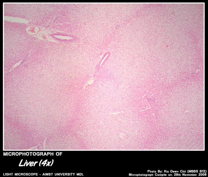
- Made up of hexagonal areas – hepatic lobules.
- Each separated – connective tissue septa.
- Each lobule has cords of liver cells separated by sinusoids.
- Each liver cell is
- - one cell thick
- - anatomizing with one another forming a network
- - spaces between the liver cells is sinusoids
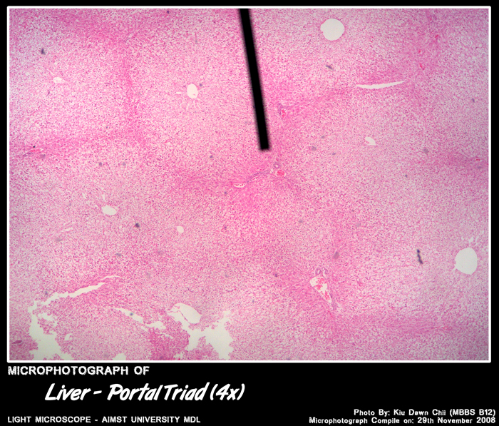
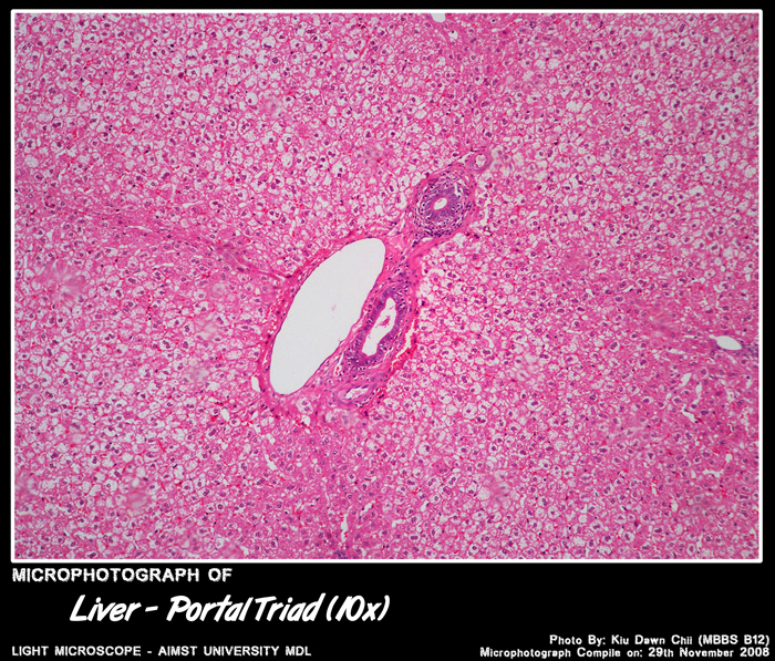
- Along the periphery of each lobule there an area filled with connective tissue.
- This area is called the portal canals.
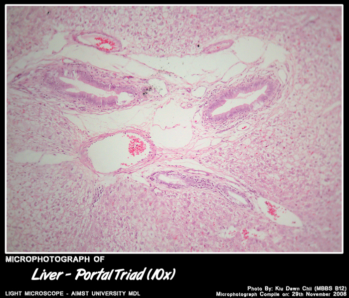
- Portal canals has
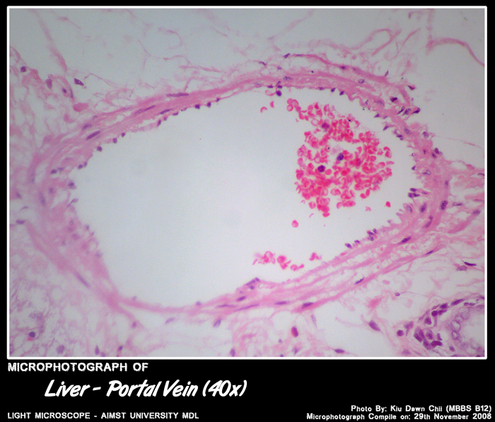
- 1. branch of portal vein
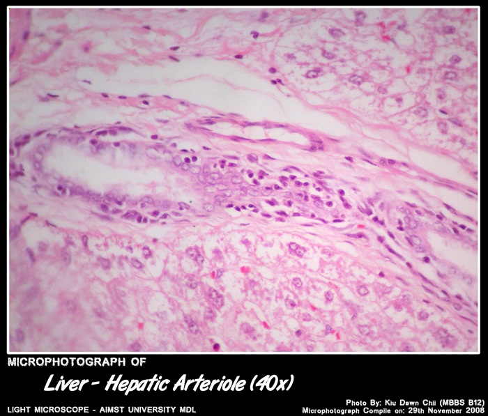
- 2. branch of hepatic artery
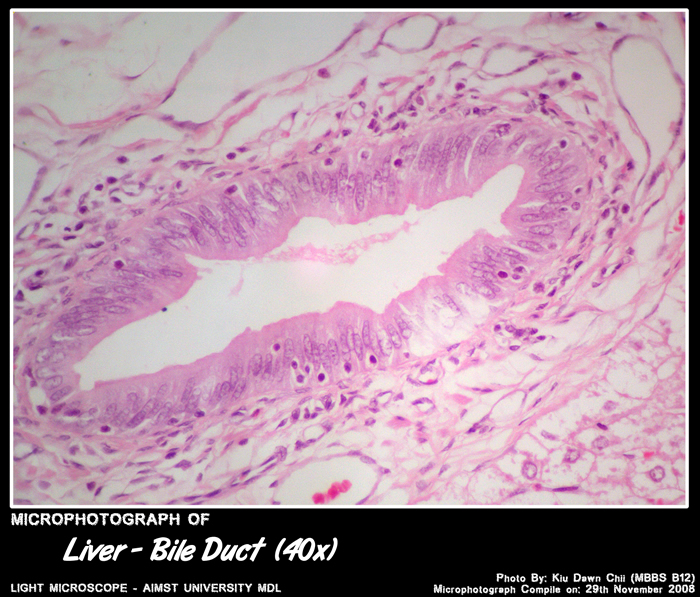
- 3. interlobular bile duct
- knowns as PORTAL TRIAD
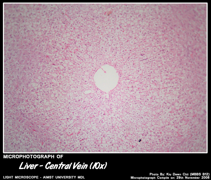
- Blood from the portal vein, hepatic artery enter the sinusoids at the periphery of the lobule.
- The blood flows towards the centre and open into a vein in the middle called central vein .
- Central veins drains into the hepatic vein – IVC
Click to enlarge